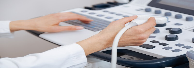
Ultrasound imaging, also called ultrasound scanning or sonography, involves the use of a small transducer (probe) and ultrasound gel to expose the body to high -frequency sound waves. Ultrasound is safe and painless, and produces pictures of the inside of the body using sound waves. Ultrasound examinations do not use Ionizing radiation (as used in x-rays). Because ultrasound images are captured in real-time, they can show the structure and movement of the body's internal organs, As well as blood flowing through blood vessels.
Conventional ultrasound displays the images in thin, flat sections of the body. Advancements in ultrasound technology include three-dimensional (3-D) ultrasound that formats the sound wave data into 3-D images. Four-dimensional (4-D) ultrasound is 3-D ultrasound in motion.
Doppler ultrasound is a special ultrasound technique that evaluates blood flow through a blood vessel, including the body's major arteries and veins in the abdomen, arms, legs and neck.
Why should I do it ?
- The most common reason for a venous ultrasound exam is to search for blood clots, especially in the veins of the leg. This condition is often referred to as deep vein thrombosis or DVT. These clots may break off and pass into the lungs, where they can cause a dangerous condition called pulmonary embolism. If the blood clot in the leg is found early enough, treatment can be started to prevent it from passing to the lung.
- A venous ultrasound study is also performed to:
- Determine the cause of long-standing leg swelling. In people with a common condition called "varicose veins", the valves that normally keep blood flowing back to the heart may be damaged, and venous ultrasound can help identify the damaged valves and abnormal blood flow.
- Aid in guiding placement of a needle or catheter into a vein. Sonography can help locate the exact site of the vein and avoid complications, such as bleeding or damage to a nearby nerve or artery.
- Map out the veins in the leg or arm so that pieces of vein may be removed and used to bypass a narrowed or blocked blood vessel. An example is using portions of vein from the leg to surgically bypass narrowed heart (coronary) arteries.
- Examine a blood vessel graft used for dialysis if it is not working as expected; for example, the graft may be narrowed or blocked.
- In children, venous ultrasound is used to:
- Evaluate a connection between an artery and a vein which can be seen in congenital vascular malformations (arteriovenous malformations or fistula) and in dialysis fistula.
- If a line is placed in a vein of the legs or arms, there is a much higher chance of developing a clot around it due to the smaller vessel size (especially in infants and young children). In some instances, a clot can suddenly form in the arm due to compression of the vein at the inlet of the chest or in the left leg due to compression of the vein on the left side by an artery in the abdomen.
- Doppler ultrasound images can help the physician to see and evaluate:
- Blockages to blood flow (such as clots).
- Narrowing of vessels
- Tumors and congenital vascular malformations
- Reduced or absent blood flow to various organs, such as the testes or ovary
- Increased blood flow, which may be a sign of infection
Any preparations needed?
- You should wear comfortable, loose-fitting clothing.
- You may need to remove all clothing and jewelry in the area to be examined.
- In case of children, ultrasound examinations are very sensitive to motion, and an active or crying child will slow the examination process. To ensure a smooth experience, it would be beneficial to explain the procedure to the child prior to the exam.
- A period of fasting is necessary only if you are to have an examination of veins in your abdomen. In this case, you will probably be asked not to ingest any food or fluids except water for six to eight hours ahead of time. Otherwise, there is no other special preparation for a venous ultrasound.